What’s the real difference between X-ray, CT and MRI?
You’d be surprised how often these three are confused — even by people who’ve had scans themselves.
As a massage therapist, I regularly hear things like, “I had a scan and they said everything was fine.”
The next question is always: what kind of scan?
Let’s break it down in a simple, friendly way.


X-ray
An X-ray is the most basic and commonly used imaging method.
It:
- Uses radiation
- Produces a flat, two-dimensional image
- Is quick and relatively inexpensive
- X-rays are excellent for showing hard structures: Broken bones, Joint alignment, Severe arthritis, Chest conditions (like pneumonia).
If you suspect a fracture, an X-ray is usually the first step.
But here’s the key point:
👉 X-rays do not show soft tissues clearly.
They won’t properly show discs, nerves, ligaments or muscles.
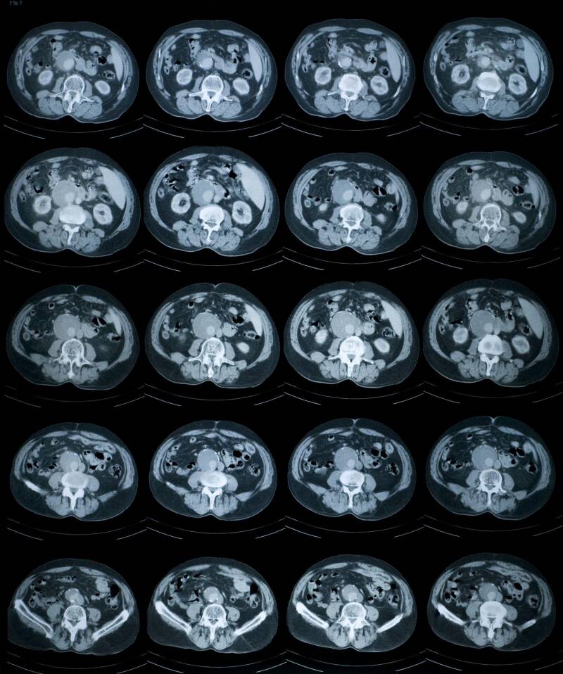
CT Scan (Computed Tomography)
A CT scan also uses X-rays — but in a much more advanced way.
Instead of a single flat image, it creates:
Layered cross-sections
- Detailed 3D-style images
- CT scans are particularly useful for: Complex fractures, Internal bleeding, Head injuries, Chest and abdominal problems.
Think of CT as a more detailed, multi-layered version of an X-ray.
It’s still mainly focused on structure, especially bone and internal organs — but it’s not the best tool for subtle soft-tissue problems in the spine.


MRI Scan (Magnetic Resonance Imaging)
MRI works completely differently.
It:
- Uses magnetic fields
- Uses no radiation
- Produces extremely detailed images of soft tissues
MRI is the gold standard for: Herniated or bulging discs, Nerve compression, Ligament injuries, Muscle tears, Brain and spinal cord conditions.
If someone has persistent back pain, sciatica or suspected disc issues, an X-ray will not reveal the real cause. An MRI is usually required.
The Simple Way to Remember It
👉 Hard structures = X-ray or CT
👉 Soft tissues = MRI
Wrong tool = wrong information.
A Real-Life Example
One client once told me she had an X-ray done at a private clinic before a spinal adjustment. She was told everything looked “perfect”.
But she was still in pain.
The issue?
X-rays don’t show discs or nerves. If the problem is a disc protrusion or nerve irritation, it simply won’t appear on that image.
It’s a bit like trying to listen to music with your TV remote — nothing is technically “broken”, you’re just using the wrong device.
Why This Matters
Choosing the correct diagnostic method is important for safety.
You wouldn’t visit a dentist for stomach pain.
In the same way, you need the right scan for the right problem.
Understanding this helps you: Ask better questions, Make informed decisions, Avoid unnecessary procedures, Protect your spine and nervous system.
If you’re ever unsure what kind of imaging is appropriate, speak to your GP or consultant first.
And if you’d like it explained in simple terms, I’m always happy to talk through it during a session. The more you understand your body, the safer and more confident you’ll feel about your health.
21 / 12 / 2025